Scientific Research
Fine Art X-Ray Research
Digital radiography for art authentication and preservation
Overview
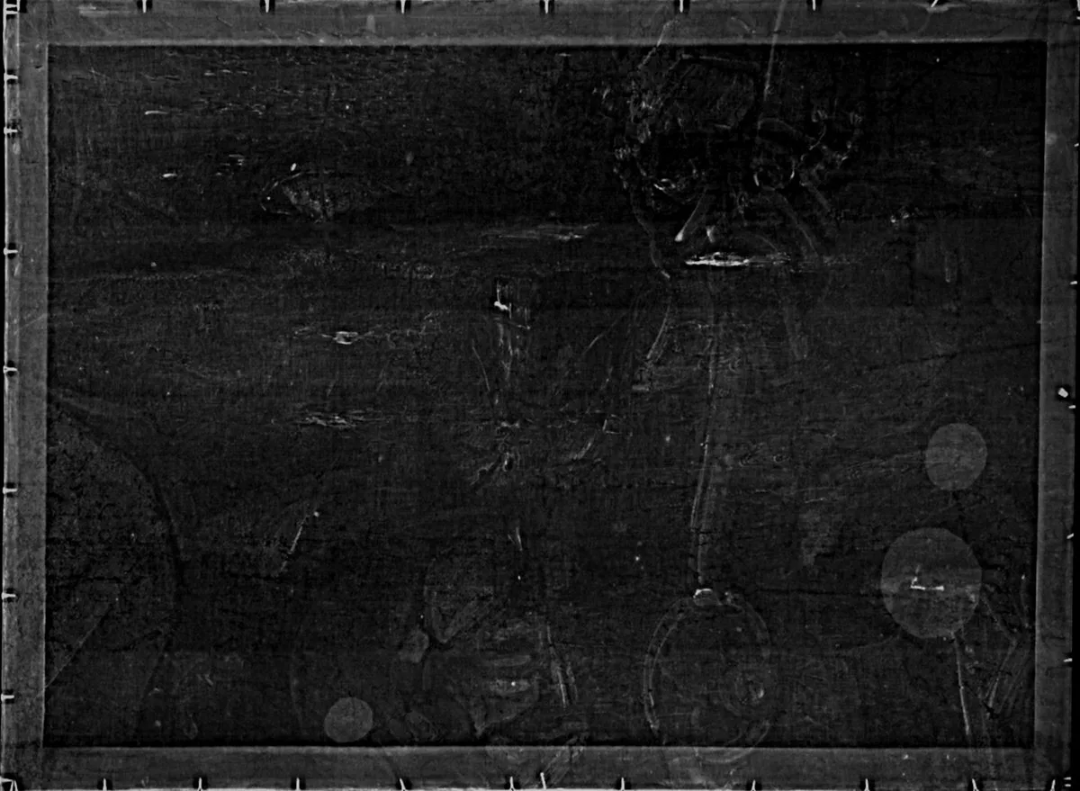
The fine art radiograph (x-ray) is used for discovery in Smith's fine art research. Digital radiography exposes the creative process and other information critical to a specific artist. The advancement of digital technology allows a painting to be scanned without the creation of a composite of many individual film x-rays.
History of the Research
The fine art digital radiography (x-ray) project started October 30, 2003 with The McCrone Associates, Inc. in Chicago, IL. The McCrone Associates, Inc. under the direction of Joseph Barabe created a composite of six digital radiographs (x-rays) of a 16"x20" painting for review by Smith. The painting was scanned using an electric sensor at a Chicago medical facility. Each scan was then produced on film.
This was Smith's first hands-on use of digital radiography. Prior, Smith gained experience reviewing film radiographs (x-rays) while working with the claims, underwriting and accounting departments of an insurance company.
Creighton University Collaboration
Smith met Dr. Roger Aiken, Director, Fine Art Department of Creighton University, Omaha, NE, USA in 2003 and agreed to be a guest speaker for his art history class. The following year Smith developed individual fine art research programs for women on the dean's list. She mentored women fine art students at Creighton University for 6 years. During these years she was a guest speaker at Creighton University for the Fine Art Department.
Creighton University fine art department introduced her to the Creighton University Medical Center (CUMC) medical director in Omaha, NE, USA. The new digital radiography technology, the LODOX Statscan, was on loan to CUMC. The CUMC board of directors approved the use of the Statscan during down hours for Smith's fine art research.
LODOX Statscan Partnership
In 2004 the LODOX Statscan fine art project began. Smith utilized the equipment for her fine art research and authentication projects at Creighton University Medical Center (CUMC), Omaha, NE facility. The inventor, developers, and LODOX staff involved with the Statscan from Johannesburg, South Africa and Painesville, Ohio visited Omaha, Nebraska for Smith's Brown Bag Lunch lecture "Art & Science" for doctors at CUMC. The lecture featured radiographs created utilizing LODOX Statscan technology used in Smith's fine art research and journal articles.
A poster of Smith's original fine art research images created with the LODOX Statscan was exhibited June 28 through July 1, 2006 at the International CARS Conference in Osaka, Japan. The published poster title was "028 Digital radiography in the Analysis of Artwork."
Over the years with the continued support of the LODOX Corporation, Smith has scanned and created radiographs (x-ray) of paintings and pictures nationwide with the Statscan technology. The technology is located throughout the USA. Smith remains independent of any institution for her fine art research and fine art x-ray projects.
Inspiration and Methodology
Smith credits the research and fine art publications of Madeleine Hours, director of the laboratory at the Louvre, for her interest in fine art forensics and x-ray. Smith thanks the Louvre for allowing women fine art majors nationwide under her direction to complete research projects over the years at the Louvre Museum Archives in Paris, FR.
Digital radiography creates a fingerprint of a painting and the artist's creative process, thereby protecting it from theft, forgery and preserving the artist's catalogue raisonne for future generations.
Acknowledgements
Thank you, appreciation and acknowledgement to the LODOX Corporation for their continued support of Smith's fine art research projects throughout the world, wherever the technology is available. The value they have added to fine art research is immeasurable.
Acknowledgement and appreciation to Creighton University Fine Art Department and Creighton University Medical Center board of directors and medical staff for their support for the fine art radiograph (x-ray) project.
Special thanks to Joe Barabe and The McCrone Associates, Inc., Chicago, IL for their fine art review assistance, pigment analysis and guidance over the years.
The Ford Conservation Center in Omaha, NE, USA is critical to many successful fine art projects with the use of their library and collaboration with their knowledgeable director and staff. Specific thanks to Julie Reilly, Executive Director.
The Great Plains Art Museum, Love Library, University of Nebraska, Lincoln; Joslyn Art Museum, Omaha, NE; Sheldon Museum of Art, University of Nebraska, Lincoln; and Museum of Nebraska Art, University of Nebraska-Kearney are valued library resources.
Special thanks to the University of Central Florida, Orlando, FL; The Nelson-Atkins Museum of Art, Kansas City, MO; Alfred R. Goldstein Library, Ringling College of Art & Design, Sarasota, FL; Museum of the North, University of Alaska, Fairbanks, AK; Indianapolis Museum of Art, Indianapolis, IN; Fogg Museum, Harvard University, Cambridge, MA; Anchorage Museum, Anchorage, AK; Smithsonian American Art Museum, Washington, DC; and The Getty Research Institute, Los Angeles, CA; and many regional fine art galleries and museums throughout the United States of America. The directors, curators and staff have been vital to years of fine art research conducted by Smith.
Smith recommends the MACC workshop, Minneapolis, MN, "Conservation Procedures that Aid in Identifying Fakes & Forgeries in the Art World."
Have a similar project? We’d love to hear from you.
Begin a Conversation